Kidney stone
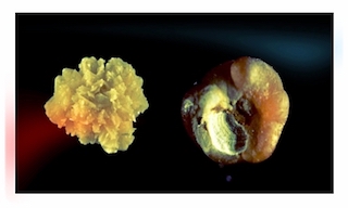
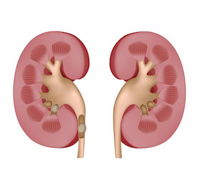
Urinary stones or urolithiasis are "stones" that form inside the urinary tract. They develop in the kidney and correspond to the slow build-up of microcrystals in urine. They can measure a few millimeters to the size of a golf ball.
10% of the population will manufacture at least one calculus and in 50% of cases these people will reoffend.
The causes are most often multiple (diet, lack of hydration, sedentary lifestyle, urinary tract infection, etc.), in patients with multiple recidivism, it is necessary to know how to look for an underlying cause (a hormonal disease, a genetic disease, etc.). Prevention also involves the analysis of the stone eliminated, or removed by surgery, in order to specify its composition (infrared analysis).
When the stones remain in the kidney they are most often asymptomatic (do not give pain), it is when they migrate into the ureter that they can then block the flow of urine between the kidney and the bladder. and cause a sudden and violent pain called renal colic crisis.
Renal colic
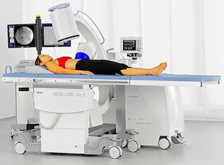
Medical treatment is still needed to calm the pain of renal colic. Sometimes we need to administer morphine products to the emergency room. It is recommended to perform a CT scan without injection of contrast product to determine the site and size of the calculus.
A stone whose size is less than 5 mm can be eliminated spontaneously but this is not always the case !. Urgent drainage is sometimes necessary if medical treatment is insufficient to relieve the pain or if there is a fever.
Minimally invasive techniques
- Shock wave lithotripsy (SWL)
- Flexible laser ureteroscopy
- Ureteroscopy
- double J Stenting
- Percutaneous nephrolithotripsy


Shock wave lithotripsy (SWL)
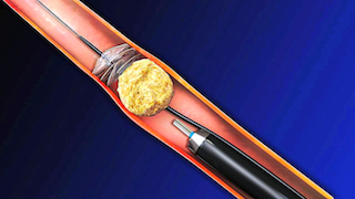
This is a system that sends shock waves through the skin into the stone, causing it to explode. This minimally invasive treatment is mainly used for kidney stones larger than 5 mm and for stones in the lower ureter. The complete success rate is in the order of 60%.
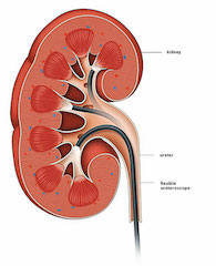
Flexible laser ureteroscopy: This new cutting-edge technique uses an endoscope (a kind of small camera through which instruments can pass: laser fiber, forceps, basket probe, etc.); this type of procedure is performed under general anesthesia and often only requires a 24-hour hospital stay. The endoscope is introduced by natural means and travels up the urinary tract until it comes into contact with the stone in the kidney. Once positioned, the laser fiber is used so as to fragment the calculation. The complete success rate is around 90%. (video)
Flexible ureteroscopy 3D
Rigid ureteroscopy: This is a rigid but very thin instrument that is used to treat stones in the ureter (urinary canal connecting the kidney to the bladder). Different instruments are used to fragment and remove the stone. A double J catheter is often put in place for a few weeks. It will then be removed under local anesthesia.
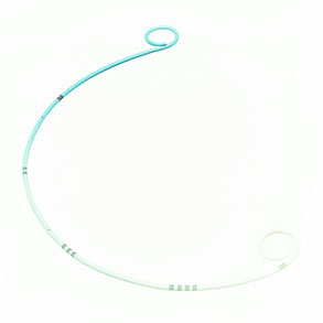
The "double J stent" is a thin hollow silicone tube with the upper end positioned in the kidney and the lower end in the bladder. This probe allows the kidney to be emptied when it is obstructed and provides rapid relief to the patient in whom medical treatment was insufficient. This catheter can cause frequent urges to urinate. It is removed after a few weeks under local anesthesia and does not require further hospitalization.
PCNL
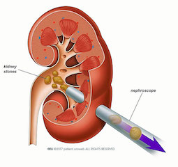
Percutaneous nephrolithotripsy (PCNL) is used to treat large stones, this intervention takes place under general anesthesia; it consists of putting a rigid tube through the skin of the back into the kidney, once in place, this access is used to introduce an optical system coupled to an instrument that crushes the calculus, or a laser. The fragments are then removed by specific forceps. The patient will keep a small scar that does not exceed 2 cm.
Cabinet d'urologie
Dr Wassim Chaabane
Centre Médical Coral
Centre Urbain Nord, 1082 Tunis
كورال الطبي
المركز العمراني الشمالي تونس
+216 71 947 574

Traitements innovants
Enucleation Prostatique au plasma
Laser pour calcul
micro-TESE
Varicocèle
Contact
Téléconsultation
A Propos
